Free Microscope Drawing, Download Free Microscope Drawing png images
Parts Of a microscope. The main parts of a microscope that are easy to identify include: Head: The upper part of the microscope that houses the optical elements of the unit.; Base: The base is attached to a frame (arm) that is connected to the head of the device.The base of the microscope provides stability to the device and allows the user's hands to be free to manipulate other aspects of.

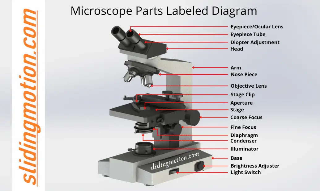
Parts of a microscope with functions and labeled diagram
microscope, instrument that produces enlarged images of small objects, allowing the observer an exceedingly close view of minute structures at a scale convenient for examination and analysis.Although optical microscopes are the subject of this article, an image may also be enlarged by many other wave forms, including acoustic, X-ray, or electron beam, and be received by direct or digital.

36 Label Parts Of The Microscope Labels 2021
The parts of the compound microscope is categorized into two - the mechanical parts and the optical parts. It is also known as bright-field microscope because it enables the light to pass directly through the source of light through the two lenses. Let us discuss the different parts of a compound microscope. a.
How to Use a Microscope
These labeled microscope diagrams and the functions of its various parts, attempt to simplify the microscope for you. However, as the saying goes, 'practice makes perfect', here is a blank compound microscope diagram and blank electron microscope diagram to label. Download the diagrams and practice labeling the different parts of these.

Microscope Parts Worksheet Answer Key Thekidsworksheet
A labeled diagram of microscope parts furnishes comprehensive information regarding their composition and spatial arrangement within the microscope, enabling researchers to comprehend their function effectively. In this comprehensive article, we will delve into the intricate parts of the microscope, exploring their functions in detail.

Microscope parts, Microscope, Anatomy and physiology book
This part is where doctors or scientists put the microscope slide with samples for analysis; it normally has clips that prevents a slide from moving while it is being viewed by the user from the eyepiece part; the slide can be moved manually while it is being viewed or it can be moved mechanically if you are using a microscope with a mechanical platform; this is done by rotating the knobs.

🎉 Main components of a light microscope. Parts of a microscope with
The common light microscope used in the laboratory is called a compound microscope. It is because it contains two types of lenses; ocular and objective. The ocular lens is the lens close to the eye, and the objective lens is the lens close to the object. These lenses work together to magnify the image of an object. Parts of Compound Microscope

Microscope Diagram Labeled, Unlabeled and Blank Parts of a Microscope
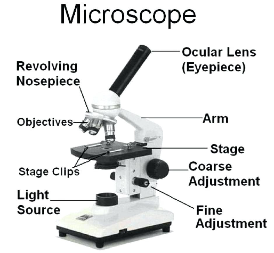
The individual parts of a compound microscope can vary heavily depending on the configuration & applications that the scope is being used for. Common compound microscope parts include: Compound Microscope Definitions for Labels Eyepiece (ocular lens) with or without Pointer: The part that is looked through at the top of the compound microscope.
Parts Parts And Functions Of A Microscope
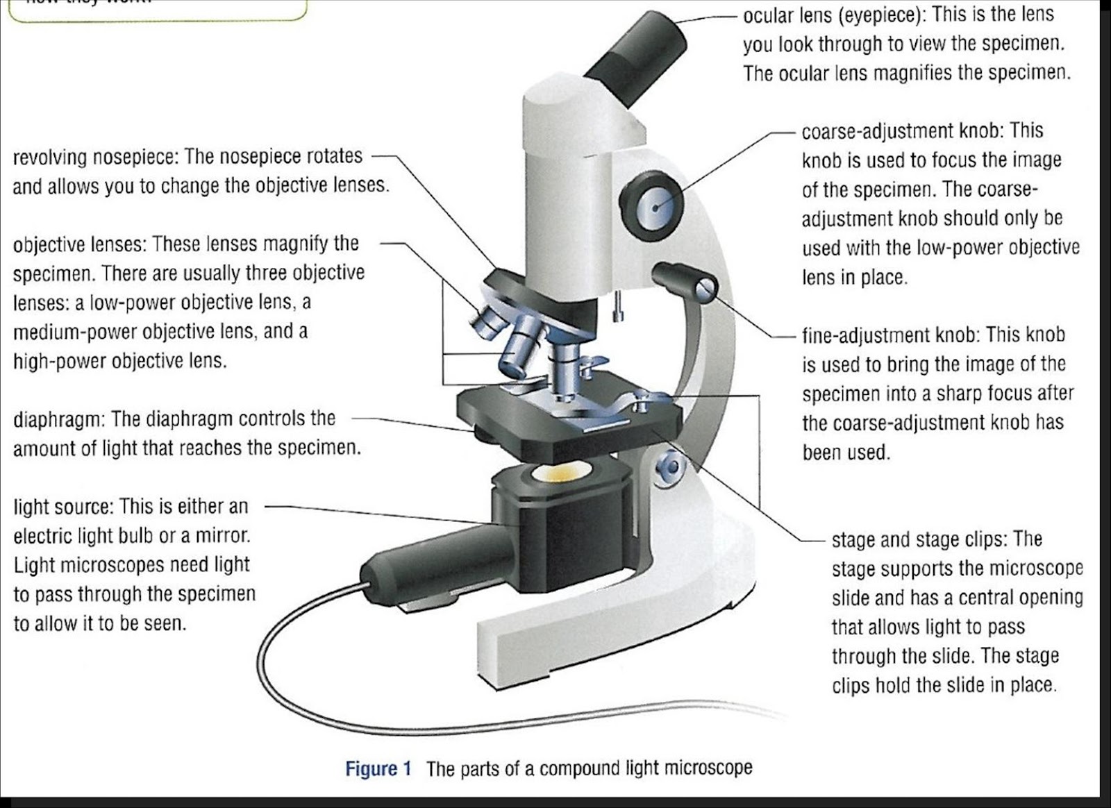
Figure 2: Brightfield light microscope used in a Microbiology lab (Lumen) The Optical System. The optical system of a compound microscope consists of two lens systems: one found in the objective(s) lens(es) (Fig. 2, part 3); the other in the ocular (eyepiece) (Fig. 2 part 1).

301 Moved Permanently
Explore the different parts of a microscope using a diagram, including the microscope lens, eyepiece, and stage. Updated: 10/13/2022 Create an account Table of Contents. What is a Microscope?.

Diagrams of a Microscope 101 Diagrams
A microscope is an instrument that magnifies objects otherwise too small to be seen, producing an image in which the object appears larger.. In most cases, the part of a cell or tissue that we want to look at isn't naturally fluorescent, and instead must be labeled with a fluorescent dye or tag before it goes on the microscope..

Academic Biology
Microscope Parts & Specifications. Historians credit the invention of the compound microscope to the Dutch spectacle maker, Zacharias Janssen, around the year 1590 (more history here).The compound microscope uses lenses and light to enlarge the image and is also called an optical or light microscope (versus an electron microscope).
Microscope Parts Sketch at Explore collection of
Parts of Compound Microscope/ Light microscope. Eyepiece (Ocular): it is the lens through which we observe the magnified picture. It enlarges the image created by the objective lenses and gives a more pleasant viewing experience. The eyepiece's principal purpose is to concentrate light rays and communicate the enlarged picture to the viewer.

Microscope Diagram to Print 101 Diagrams
2. Compound Microscope. Compound Microscope is a type of microscope that used visible light for illumination and multiple lenses system for magnification of specimen. Generally, it consists of two lenses; objective lens and ocular lens. It can magnify images up to 1000X.

Microscope Diagram to Print 101 Diagrams
The microscope illustrated in Figure 5 below was manufactured by Hugh Powell and Peter Lealand around 1850. The tripod base provided a sturdy support for the microscope, which many people consider the most advanced of its period. Parts of a Powell and Leland Microscope Diagram
Guide to understand microscope parts, names, functions & diagram
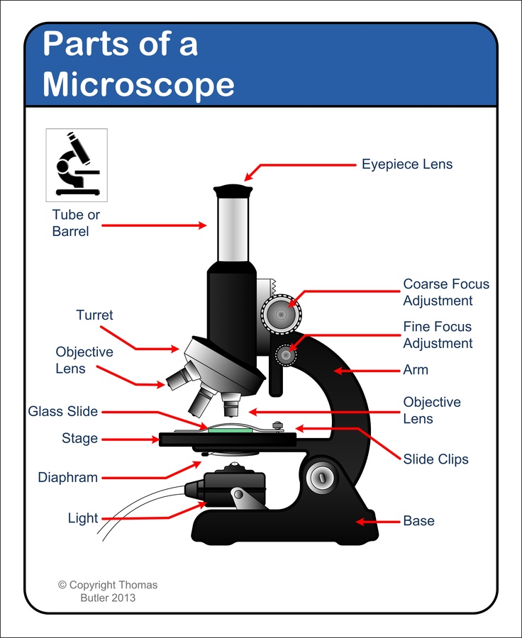
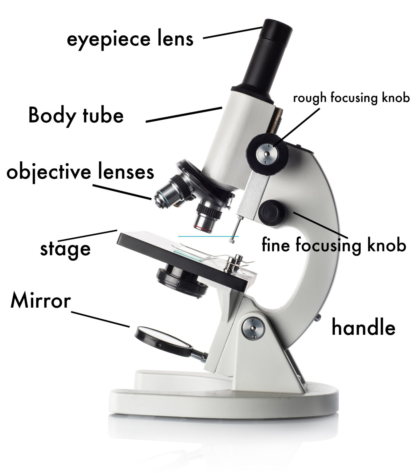
Structural parts of a microscope and their functions Figure: Diagram of parts of a microscope. There are three structural parts of the microscope i.e. head, arm, and base. Head - The head is a cylindrical metallic tube that holds the eyepiece lens at one end and connects to the nose piece at other end. It is also called a body tube or.